

The content is published under Creative Commons Attribution 4.0 International license.
Reviewed Article:
Certain Small Contrivances. Recreating an Intestinal Condom Recipe to Determine the Potential Effects of Manufacturing on Zooarchaeology by Mass Spectrometry (ZooMS)
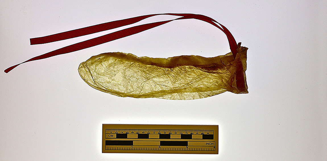
Skin and membranous artifacts are rarely recovered from archaeological excavations due to taphonomic processes that result in rapid decomposition. These classes of artifacts can, however, occasionally be preserved in extreme conditions such as waterlogging, freezing, and dry environments. One such artifact is likely an intestinal condom recovered from a well at the Oxon Hill/Addison Plantation site (18PR175; ca. 1685-early 20th c.) in Maryland, United States. To determine the taxonomic identity of this artifact, we previously employed three different Zooarchaeology by Mass Spectrometry (ZooMS) sampling techniques. While we were able to confirm that the artifact was made from a sheep, only one of our samples yielded a result. In this paper, we discuss the history of membranous condoms, provide an overview of their manufacture, and consider whether the manufacturing process could contribute to a loss of collagen yield. We conclude with recommendations for future ZooMS analysis of intestinal objects.
Introduction
Although rare, skin and membranous artifacts can occasionally be recovered under exceptional conditions, such as in waterlogged, frozen, or dry environments. These artifacts provide insight into textile and hide industries that might not leave other forms of evidence and shed light on how different species were utilized for non-dietary purposes (Kirby, et al., 2013; Brandt, et al., 2014; Fiddyment, et al., 2015; Ebsen, et al., 2019; McGrath, et al., 2019). As with other membranous artifacts, it is exceedingly rare to recover and identify an intestinal condom archaeologically. While characteristics such as shape and texture may reinforce an identification, the animal species used is also important. The caecum, a small pouch which is naturally sealed on one end, lies at the juncture between the animals' large and small intestines. In ruminants, the caecum ferments food after it passes through the rumen, enabling the animal to absorb additional nutrients (Zou, et al., 2020). Sheep caeca were commonly used for condom manufacture because they were naturally the appropriate size for this purpose.
The earliest use of the condom is shrouded in myth, but the use of barrier methods for both contraception and disease prevention is clearly quite old. Some scholars have argued that the story of King Minos of Crete represents condom use (Khan, et al., 2013; Collier, 2007). His semen was said to contain scorpions and snakes that killed anybody he had sex with, so his wife, Pasiphae, placed a goat's bladder in her vagina to capture these creatures. Additionally, oiled silk condoms were used in China by the 10th century BCE, while condoms made of tortoise shell or animal horn were utilized in Japan (Riddle and Estes, 1992; Maatouk and Moutran, 2013; Chowdhry, et al., 2019). The latter, known as kabuto-gata (helmet shape), were also used as dildos due to its rigidity. The Dutch likely introduced condoms of thin leather, known as kawa-gata (leather type)or mara-bukuro (penis sack), to Japan around the late 15th century, although women were said to prefer the stiffness of the kabuto-gata (Collier, 2007; Maatouk and Moutran, 2013).
Al-Akhawayni Bukhari, a 10th-century Persian physician, discussed the human reproductive system and various contraceptive methods at length in his book "Hidayat al-Muta'Allemin Fi al-Tibb" (Learner's Guide to Medicine) (Yarmohammadi, et al., 2013). In one proposed method, the woman should attempt to remove semen from her vagina manually or by jumping. He also suggested a variety of spermicides, oral medications, herbal fumigation, and coitus interruptus. Finally, he proposed two separate barrier methods, one of which involved inserting oak sap into the vagina. For the second, he stated: "Men should cover the penis using the animal's gallbladder and then fix this sheath to the penis, so after ejaculation, the semen pours into this bag, not into the womb" (Yarmohammadi, 2013, p.438).
In 1564, Gabriele Fallopio advocated wrapping the penis in a linen sheath secured with a ribbon. The sheath was soaked in medicinal herbs and designed to protect the wearer from syphilis. He claimed that 1100 men tried the device, and none became infected (Collier, 2007; Chowdhry, et al., 2019). However, the device remained unnamed. While some have suggested the term "condom" comes from a 17th-century Dr. (or Colonel) Condom who allegedly provided a condom to Charles II of England so the king could avoid fathering illegitimate children, there is no contemporary documentation for this individual and he may not have existed (Gaimster, et al., 1996; Moore, 2008). Others have proposed the Persian term kendu or kondu referring to a bag-like storage vessel made from animal intestines, or various Latin terms including cunnus [the female genitals] and dum [ability to function]; condĕre [protect, sheath]; or condus [receptacle] (Amy and Thiery, 2015; Chowdhry, et al., 2019).
Regardless of its origin, there is widespread evidence for condom use in Europe by the 17th century. Early condoms were made of fish bladders as well as the caeca of sheep, calves, and goats, although sheep caeca eventually became the most popular raw material. The earliest archaeologically recovered condoms, identified in fill dating to 1647 from the keep garderobe in Dudley Castle, West Midlands, England, were made from sheep or pig intestine based on microscopic analysis (Gaimster, et al., 1996). Ten condoms were recovered, five of which were presumably unused as they were nested inside one another, possibly for storage. The other five may have been used.
The process for making intestinal condoms, outlined below, took place over several days and was laborious. Given the time required, as well as an individual animal's ability to produce only one condom by virtue of possessing a single caecum, it has been inferred that intestinal condoms were likely considered luxury objects throughout the 17th, 18th, and early 19th centuries (Gaimster, et al., 1996). Despite the inferred cost, intestinal condoms appear to have been popular in Europe, with specialty stores in England and France during the 18th century. Effusive and creative praise for condoms as both a contraceptive and disease preventative can also be found in multiple British and French publications by various authors of the same period (Gaimster, et al., 1996).
While the bespoke condoms sold at specialty shops likely would have been expensive, Malthusians seeking to reinforce methods of population control distributed pamphlets containing recipes, or "receipts," for making condoms in the 19th century. They were written in plain, accessible language and distributed to the English poor. These recipes discussed everything from acquiring the proper intestines from the local butcher to sewing the ribbon onto the finished product. The recipes have slight differences between them but are consistent overall (Collier, 2007, p.126).
Although we know that sheepskin condoms were popular in 18th century Europe (Trumbach, 1998; Drucker, 2020), there is not much evidence for their production in North America until the end of the 18th century. By that point, though, one could purchase condoms from urban centers such as New York and Philadelphia. Mederic Louis Elie Moreau de St. Mery was a Frenchman operating a bookstore that specialized in "minute books" in 1794, located on Front and Walnut Streets in Philadelphia. He also imported condoms from Paris to sell out of his storefront. He wrote:
"I did not wish to deprive my business of a profitable item, the lack of which in hot climates would not, I think, be without danger. Consequently, when my old colleague and friend, Barrister Geanty, a refugee from Cape Francois in Baltimore, who had a wide knowledge of medical supplies, offered me a stock of certain small contrivances -ingenious things said to have been suggested by the stork-I agreed. I wish to say that I carried a complete assortment of them for four years; and while they were primarily intended for the use of French colonials, they were in great demand among Americans, in spite of the false shame so prevalent among the latter. Thus the use of this medium on the vast American continent dates from this time. People from San Domingo as well as from other colonies had frequent recourse for our stock,"
(Collier, 2007, p.118).
Despite de St. Mery's assertions of "false shame" among Americans, there is limited evidence for earlier condom use in North America (Brown, 1996, pp. 332-333).
Archaeological context
Recently, an archaeologically recovered condom was identified at Oxon Hill/Addison Plantation (18PR175; hereafter referred to as Oxon Hill), Maryland, United States, recovered from a well excavated in 1985 (Bowden-Gray, et al., forthcoming). Oxon Hill was established by John Addison, a wealthy English merchant, between 1685 and 1689. The Manor house itself was not built until 1710 by Addison's son, Col. Thomas Addison. The Addison Family continued to possess the property until 1810, when the land was sold to the Berry Family. No major colonial population centers were located near Oxon Hill when it was first built; however, both Washington, D.C. (1790) and Baltimore, Maryland (1729) were later established nearby. Although earlier Indigenous artifacts were identified, no pre-contact features were found in situ, and only one ceramic sherd dating to the Late Woodland was identified. All of the features excavated date to the 18th and/or 19th centuries (Garrow and Wheaton, Jr., 1986).
The well has an overall mean ceramic date of 1753.75, which was calculated by combining ceramics from all levels of the feature. At least 13m deep, it was excavated in 76 levels, which were later organized into four sections that represent distinct fill episodes. Section A consisted of the top 35 levels, characterised by a high concentration of brick, mortar, roofing slate, and blackened soil, indicating rubble from the burning of the main house. Levels 36-49 were designated Section B and consisted of preserved wood fragments, an increased quantity of artifacts and faunal material, and some brick fragments. Levels 50-57 were designated Section C, containing more organic soil, a decrease in wood fragments, and an increase in artifacts and faunal materials. Excavators reached the water table at Level 57, causing materials from upper levels to erode into the lower levels. It is possible that materials from Level 57 may have come from further up the well shaft. Finally, Levels 58-76 were designated Section D. Because this section was below the water table, preservation of organic materials was excellent. This level did not contain soil but instead had a mix of pine straw, straw, horse manure, and grass clippings. There was also a series of vertical planks found at about Level 57, which were believed to help maintain water quality (Garrow and Wheaton, Jr., 1986, pp.214-217).
The distinct fill episodes indicate that the well likely became useless as a water source during the early eighteenth century and was subsequently used for trash disposal. The artifact in question was recovered alongside other organic materials in levels 60-62 within Section D, which dates to c. 1720-1750 (Garrow and Wheaton, Jr., 1986, pp.214-217). However, it was not identified until Sara Rivers Cofield of the Maryland Archaeological Conservation (MAC) Lab recognized its potential use while looking through the collection to pull artifacts for an Outlander-themed exhibit. Working with the MAC Lab, we used Zooarchaeology by Mass Spectrometry (ZooMS) to identify which species was used to make this artifact and test the interpretation that it is a condom (Bowden-Gray, et al., forthcoming).
ZooMS is a collagen peptide mass fingerprinting technique that allows for taxonomic identification of specimens through enzyme-digested Type I collagen. Type I collagen is the most abundant and important protein in animals (Brown, et al., 2021; Richter et al., 2022). In Type I collagen, a repeating pattern of the three amino acids, Glycine (Gly)-Proline (Pro)-Hydroxyproline (Hyp), creates a compact structure that can survive well in archaeological contexts in various depositional environments (Brown, et al., 2021). In most cases, Type I collagen has also been shown to be more resilient in preservation than DNA (Buckley, et al., 2009; 2014). In tetrapods, the collagen I triple helix comprises two identical α1 chains and one α2 chain. Collagen α-chains vary greatly among animals of different clades and are important for taxonomic identifications. The slight differences in amino acid sequences allow for taxonomic differentiation between different taxa of animals. After a sample is digested by an enzyme, which cleaves the peptides at known locations, peptides are identified by differences in their masses and compared with the masses of known animal taxa.
ZooMS has been previously used on bone and skin artifacts with great success (Buckley, et al., 2009; 2010; Richter, et al., 2011; Kirby, et al., 2013; Brandt, et al., 2014; Fiddyment, et al., 2015; Ebsen, et al., 2019; McGrath, et al., 2019; Janzen, et al., 2021; Harvey, et al., 2022). While the resolution of DNA analyses is generally higher, ZooMS analyses often provide sufficient taxonomic resolutions for many archaeological questions while also being more accessible and cost-effective for researchers (Richter, et al., 2022). Of particular interest to this project were the ZooMS studies that have had success with non-destructive sampling strategies (Brandt, et al., 2014; Fiddyment, et al., 2015; McGrath, et al., 2019; Richter, et al., 2022). Prior to these studies, most, if not all, ZooMS sampling required a small fragment of bone or skin to be destructively analysed. However, many non-destructive sampling techniques rely on the triboelectric effect, where friction between plastic polymers and a protein transfers an electric charge that captures proteins on the surface of the artifact (Richter, et al., 2022). The first of the studies to utilize this method was done on uterine vellum artifacts using a PVC eraser to drag loose electrons from the collagen to the eraser (Fiddyment, et al., 2015). The triboelectric effect has also successfully been used to obtain collagen from empty plastic sample bags and vials that previously held artifacts (McGrath, et al., 2019).
For the Oxon Hill artifact, we used three sampling techniques to identify the taxon of the intestinal condom. The "original bag method" is a virtually non-destructive sampling technique that extracts collagen from the interior surface of the archival storage bag which held the sample (McGrath, et al., 2019). The "eraser method" entails gently rubbing an eraser along the artifact's surface to recover loose collagen peptides through triboelectric charge (Kirby, et al., 2013). Finally, we removed a small sample of the artifact for traditional destructive analysis (Ebsen, et al., 2019). The sampling and preparation of the Oxon Hill samples took place at the Stable Isotope and Zooarchaeology Lab at the University of Tennessee - Knoxville, and the samples were run through the Matrix-Assisted Laser Desorption/Ionization Time-of-Flight (MALDI-TOF) mass spectrometer at the University of Florida. The "destructive" and "eraser" methods yielded unusable results, while the "original bag" method yielded the only usable spectra (Buckley, et al., 2019; Buckley, et al., 2010; Buckley and Collins, 2011; Buckley, et al., 2017; Kirby, et al., 2013; Welker, et al., 2016). However, the fragmentary nature of the artifact meant that small fragments of the condom were left behind in the original bag, which may have yielded a larger overall "destructive" sample volume than the small crumb-sized piece that was removed for the intentional destructive analysis.
Our sample in the previous study produced conclusive results that the artifact from Oxon Hill was manufactured from a sheep caecum. Combined with the shape (including an intact tip) and surface texture of the object, the ZooMS results reinforce the identification of this object as a condom. However, the eraser method failed because it only analysed the surface of the artifact, and the destructive method was either drawn from too small a sample or was over-filtered during the pretreatment process (Guiry, Szpak and Richards, 2015). It is also possible that non-destructive methods failed because they only analyse the surface of the artifact, which is more susceptible to contamination or degradation. There are a variety of factors that may have influenced collagen preservation, or the object may have come into contact with other sources of collagen during use, deposition, excavation, or curation. While we cannot assess all factors potentially affecting collagen preservation, this study analyses the potential impacts of the manufacturing process on collagen yield.
Experimental Methods
| Manufacturing Stage | Description |
| Stage 1 | Separated caecum from intestines |
| Stage 2 | Caecum soaked in water overnight |
| Stage 3 | Caecum soaked in calcium hydroxide for 24 hours |
| Stage 4 | Mucous membrane removed |
| Stage 5 | Caecum exposed to vapor of sulfur |
| Stage 6 | Rinsed with castile soap and water |
| Stage 7 | Stretched over glass mold greased with olive oil, external side in |
Table 1. Manufacturing stages to produce an intestinal condom. We took samples for ZooMS analysis at the conclusion of each manufacturing stage.
To determine whether the manufacturing process would affect collagen yield, we created an intestinal condom from a sheep caecum and took samples for ZooMS analysis after each manufacturing step (See Table 1). We drew upon an 1824 recipe for making condoms:
"1824 Ordinary condoms are made from sheeps intestinal caeca soaked in water for some hours, turned inside out, macerated again in weak alkaline changed every 12 hours, scraped carefully to remove the mucous membrane, leaving the peritoneal and muscular coats; exposed to the vapour of burning brimstone, washed with soap and water; blown up, dried, cut to length of 7-8 inches, bordered at the open end with a riband.
Baudruches fines; soaked in weak ley, turned inside out, dressed as before. Soaked in ley again, brimstoned, drawn smooth upon oiled moulds of a proper size, with the external coast of the gut next to the mould.
Baudruches superfines; washed in 2 soapy waters after soaking in them for 24 hours and very carefully dressed with the knife. Soaked in hard water for 3 days, the water being often changed; dried with a clean cloth, scented with essences, and being stretched on a glass mould, rubbed with a glass to polish them. Condoms should be soaked in water before use to make them supple,"
(Collier, 2007, p.127).
The baudruches fines [fine rubber skin] were of a higher-than-normal quality, and the baudruches superfines were of the highest quality.
We created a baudruches fines condom since that also included the steps for an "ordinary" condom. To make the condom, we first acquired sheep intestines from a custom slaughter butcher in Knoxville, Tennessee. Some companies still make intestinal condoms today, and we reached out to a manufacturer to get more details regarding the breed, age, and size of sheep used for condom production to refine our selection. However, they could not share that information with us (Trojan, pers. comm., 2023). Given that calves and goats were occasionally used to produce condoms, the caecum has to be stretched over a mold to produce the condom, and a ribbon was used to secure the condom into place, there is likely a small range of acceptable sizes for condom manufacture (Collier, 2007, p.122). Therefore, we left our request with the custom slaughter butcher open to sheep of any age and size; the intestines received came from a young adult.
Once we retrieved the intestines from the butcher, we identified and removed the caecum, which had to be emptied of stool and rinsed (See Figures 1, 2). We placed the caecum in distilled water at 20:30 on Day 1 and removed it from the water at 08:10 on Day 2 (See Figure 3). We then turned the caecum inside out and created "limewater" using food-grade Calcium Hydroxide (1g Ca(OH)2 in 630 mL of H2O) to serve as our weak alkaline. We soaked the caecum in the calcium hydroxide solution for 12 hours, removed the caecum, and soaked it for an additional 12 hours in a fresh calcium hydroxide solution.
Following this step, we had to scrape "carefully to remove the mucous membrane, leaving the peritoneal and muscular coats" (Collier, 2007, p.127). While this recipe does not say what was used to remove the mucosa, another recipe from 1844 says to "remove the mucous membrane with the nail" (Collier, 2007, p.127). This could refer to one's fingernails, but we wore gloves and other PPE throughout the process and could not use our fingernails. We decided instead to use a replica machine-cut nail to scrape the mucosa. Not only does this test the other potential interpretation, scraping the mucosa with a nail fastener instead of a fingernail, but we determined that it would be a closer approximation to scraping the mucosa with a fingernail than using a sharper implement such as a knife or scalpel.
After removing the mucosa, we burned 99% pure elemental sulfur in a stainless-steel pot within a fume hood, hanging the caecum over the handle of the stainless-steel pot to expose it to the "vapour of burning brimstone." We then washed the caecum using Dr. Bronner's Unscented Pure-Castile Soap and distilled water. Finally, we stretched the caecum, external side in, over a glass mold that was oiled with olive oil (See Figure 4). We then removed the caecum from the mold, cut it to size, and sewed a ribbon around the opening (See Figure 5).
ZooMS Methods
Approximately 1-2.5 g of intestine was sampled after each stage in the manufacturing process. Seven intestinal samples were freeze-dried to remove moisture, allowing us to record an accurate starting dry weight of each sample (Brock, et al., 2010, p.106), and two 1-11 mg fragments from each sample were removed for ZooMS analysis. For each manufacturing step, one of the two samples taken was rinsed in 200 µL of 0.1M NaOH to remove non-collagenous proteins that could affect the resulting spectra (Ebsen, et al., 2019; Lee, et al., 2022; Matinong, et al., 2022). NaOH causes the treated matrix to swell due to the breakdown of intermolecular bonds within the protein, which converts insoluble proteins into soluble proteins, resulting in increased protein hydrolysis. The increased protein hydrolysis enhances the removal of proteins but does not cause collagen hydrolysis because of collagen's triple helical structure. The removal of non-collagenous proteins using a NaOH treatment increases collagen content and purity (Lee, et al., 2022; Matinong, et al., 2022). The remaining samples were immediately rinsed with ammonium bicarbonate (AmBic). These two pretreatment methods were utilized to determine if non-collagenous proteins were obscuring ZooMS peptide peaks.
The samples were then rinsed two times with ultra-pure water and one time with 50 µL of AmBic, then suspended in 50 µL of AmBic. All samples were digested with 1 µL of 0.4 µg/µl trypsin solution and incubated at 37˚C for 6 hours. Digestion was terminated with the addition of 1 µL of 5% (v/v) trifluoroacetic acid (TFA), and samples were examined directly without further purification (Ebsen, et al., 2019; Brown, et al., 2020). Peptides were then spotted in duplicate onto a MALDI plate with a matrix, α-cyano-4-hydroxycinnamic acid (ACHA) that co-crystalizes with the peptides (Richter, et al., 2022). Samples were prepared at the University of Tennessee's Stable Isotope and Zooarchaeology Laboratory in the Department of Anthropology. After spotting, the MALDI plate was sent to Harvard University for MALDI-TOF analysis. Mass spectra were acquired for the mass to charge ratio (m/z) range 800-3500 Da.
Spectral analysis was performed using the open-source software mMass. Each spectrum was inspected manually for peptide markers A-G and markers P1 and P2 (Buckley, et al., 2009; Kirby, et al. 2013; Buckley, et al., 2014; Buckley, et al., 2017; Ebsen, et al., 2019). Spectra of poor quality were designated as not identifiable or NID. Poor quality samples were defined as those with low signal to noise (S/N) ratios and those that showed no distinct marker. Peptide markers were compared to previously published reference markers for sheep (Ovis aries) (Buckley, et al., 2009; Buckley, et al., 2010; Buckley and Collins, 2011; Kirby, et al., 2013; Welker, et al., 2016; Buckley, Harvey and Chamberlain, 2017).
ZooMS Results
For each manufacturing stage, we ran two samples: one pre-treated with NaOH, and one that was not pre-treated. Of the 14 samples analysed, three (21.4%) yielded non-identifiable spectra. The remaining 11 samples (78.6%) resulted in clear species identification as sheep (See Graphs 1, 2). All pre-treated samples yielded clear spectra for interpretation, whereas three of the seven that were not pre-treated with NaOH yielded non-identifiable spectra (See Graphs 3, 4).
The composition and structure of intestinal tissues vary among different species, with some animals, such as sheep, having a higher fat content. Treatment with NaOH can also serve to remove excess lipids that may overload the sample (Matinong, et al., 2022). Spectra were consistent across all pre-treated samples, indicating that no steps in the condom manufacturing process affected the recovery of collagen for ZooMS. Instead, it is likely that much of the collagen degradation that occurs within archaeological membranous artifacts is taphonomic in nature.
Conclusion
This study suggests that neither chemical nor physical manufacturing processes significantly affect collagen yield. Instead, taphonomic processes are the most likely cause of collagen deterioration for intestinal artifacts. McGrath et al. (2019) found that peak intensity tends to be lower using non-destructive techniques. High molecular weight peptides, often those that are used to discriminate between closely related species, are missing from many of the results of non-destructive sampling techniques. The absence of these high-weight peptides is due to the breakage of longer peptides during the removal of the collagen from the surface of the bag or artifact (McGrath, et al., 2019). Damage to collagen strands through the sample collection process or sample degradation via environmental and age factors are the most likely reason for unclear non-destructive sample results. Although lower collagen yields may cause non-destructive sampling methods to fail, utilizing the "original bag" method of ZooMS sampling was still effective in determining that the Oxon Hill artifact was manufactured from a sheep. This is likely due to the fact that the original artifact was "crumbly," leaving behind small fragments that could not be removed from the original bag. Combined with microscopic analysis and the intact tip of the object, we were able to confidently identify this artifact as a condom. ZooMS continues to be efficacious in identifying leather, skin, and membranous materials (Kirby, et al., 2013; Brandt, et al., 2014; Fiddyment, et al., 2015; Ebsen, et al., 2019); therefore, we recommend ZooMS with NaOH pretreatment for identifying archaeological intestinal artifacts in future studies.
Acknowledgements
We would like to acknowledge and thank EXARC for their encouragement and financial support for this project through the Kiernan Experimental Archaeology Award. We are very grateful to Anneke Janzen and Barbara Heath from the University of Tennessee - Knoxville for the use of university lab spaces to conduct this research and for their commentary on earlier versions of this publication. We are grateful to Christine Korzow Richter, formerly at Harvard University, now at Texas A&M University, for running our experimental samples through the MALDI-TOF and John Krigbaum at the University of Florida for running our archaeological samples through the MALDI-TOF. We are also grateful to Jenna Watson for assisting in the dissection and removal of the caecum from the intestines of the sheep. Finally, we would like to thank Rebecca Webster, Jonah Bullen, Kandace Hollenbach, Sara Rivers Cofield, the Maryland Archaeological Conservation Laboratory (MAC Lab), and the Gloria King Fellowship for their support of the Oxon Hill Condom Project that inspired this research.
Keywords
Country
- USA
Bibliography
Amy, J.J. and Thiery, M., 2015. The condom: a turbulent history. The European Journal of Contraception and Reproductive Health Care, 20(5), pp.387-402.
Bowden-Gray, T., E.G. Tarulis, B.M. Ogden, and S. Rivers Cofield (Forthcoming) Gut Check: An Examination of an 18th-Century Intestinal Condom from Oxon Hill Manor (18PR175), Maryland, USA, using ZooMS and Isotopic Analysis [Unpublished Manuscript]. Department of Anthropology, University of Tennessee, Knoxville.
Brandt, L.Ø., Schmidt, A.L., Mannering, U., Sarret, M., Kelstrup, C.D., Olsen, J.V. and Cappellini, E., 2014. Species Identification of Archaeological Skin Objects from Danish Bogs: Comparison between Mass Spectrometry-Based Peptide Sequencing and Microscopy-Based Methods. PLoS ONE, 9(9), e106875.
Brock, F., Higham, T., Ditchfield, P. and Ramsey, C.B., 2010. Current Pretreatment Methods for AMS Radiocarbon Dating at the Oxford Radiocarbon Accelerator Unit (ORAU). Radiocarbon 52(1), pp.103-112.
Brown, K.M., 1996. Good wives, nasty wenches, and anxious patriarchs: gender, race, and power in colonial Virginia. Chapel Hill, North Carolina: Omohundro Institute of Early American History and Culture and the University of North Carolina Press.
Brown, S., Heberstreit, S., Wang, N., Boivin, N., Douka, K. and Richter, K.K., 2020. Zooarchaeology by Mass Spectrometry (ZooMS)- Pretreatment protocols for bone material v1. Available at: <dx.doi.org/10.17504/protocols.io.bf5djq26>
Brown, S., Douka, K., Collins, M.J. and Richter, K.K., 2021. On the standardization of ZooMS nomenclature. Journal of Proteomics, 235, 104041.
Buckley, M., Collins, M., Thomas-Oates, J. and Wilson, J.C., 2009. Species identification by analysis of bone collagen using matrix-assisted laser desorption/ionisation time-of-flight mass spectrometry. Rapid Communications in Mass Spectrometry, 23(23), pp.3843-3854.
Buckley, M., Whitcher Kansa, S., Howard, S., Campbell, S., Thomas-Oates, J. and Collins, M., 2010. Distinguishing between archaeological sheep and goat bones using a single collagen peptide. Journal of archaeological science, 37(1), pp.13-20.
Buckley, M. and Collins, M.J., 2011. Collagen survival and its use for species identification in Holocene-lower Pleistocene bone fragments from British archaeological and paleontological sites. Antiqua, 1(1), e1-e1.
Buckley, M., Fraser, S., Herman, J., Melton, N.D., Mulville, J. and Pálsdóttir, A.H., 2014. Species Identification of Archaeological Marine Mammals Using Collagen Fingerprinting. Journal of Archaeological Science, 41, pp.631-641.
Buckley, M., Harvey, V.L. and Chamberlain, A.T., 2017. Species identification and decay assessment of Late Pleistocene fragmentary vertebrate remains from Pin Hole Cave (Creswell Crags, UK) using collagen fingerprinting. Boreas, 46(3), pp.402-411.
Chowdhry, S., Jaiswal, P., D'Souza, P. and Dhali, T.K., 2019. The Condom. Sexuality and Culture, 23(2), pp.674-683.
Collier, A., 2007. The humble little condom: a history. Amherst, New York: Prometheus Books.
Drucker, D.J., 2020. Contraception: A Concise History. Cambridge, Massachusetts: The MIT Press.
Ebsen, J.A., Haase, K., Larsen, R., Vestergaard Poulsen Sommer, D. and Brandt, L.Ø., 2019. Identifying Archaeological Leather - Discussing the Potential of Grain Pattern Analysis and Zooarchaeology by Mass Spectrometry (ZooMS) through a Case Study Involving Medieval Shoe Parts from Denmark. Journal of Cultural Heritage, 39, pp.21-31.
Fiddyment, S., Holsinger, B., Ruzzier, C. and Collins, M.J., 2015. Animal Origin of 13th-century Uterine Vellum Revealed Using Noninvasive Peptide Fingerprinting. PNAS, 112(49), pp.15066-15071.
Gaimster, D., Boland, P., Linnane, S. and Cartwright, C., 1996. The archaeology of private life: the Dudley Castle condoms. Post-Medieval Archaeology, 30(1), pp.129-142.
Garrow, P.H. and Wheaton, Jr., T.R., 1986. Final Report, Oxon Hill Manor, Archaeological Site Mitigation Project, I-95/MD 210/I-295. Vols 1 and 2. Report prepared for the Maryland Department of Transportation by Garrow and Associates, Inc.
Guiry, E.J., Szpak, P. and Richards, M.P., 2015. Effects of lipid extraction and ultrafiltration on stable carbon and nitrogen isotopic compositions of fish bone collagen. Rapid communications in mass spectrometry, 30(13), pp.1591-1600.
Harvey, V.L., LeFebvre, M.J., Sharpe, A.E., Toftgaard, C., deFrance, S.D., Giovas, C.M., Fitzpatrick, S.M. and Buckley, M., 2022. Collagen Fingerprinting of Caribbean Archaeological Fish Bones: Methodological Implications for Historical Fisheries Baselines and Anthropogenic Change. Journal of Archaeological Science, 145(September), 105642.
Janzen, A., Richter, K.K., Mwebi, O., Brown, S., Onduso, V., Gatwiri, F., Ndiema, E., Katongo, M., Goldstein, S.T., Douka, K. and Bolvin, N., 2021. Distinguishing African Bovids Using Zooarchaeology by Mass Spectrometry (ZooMS): New Peptide Markers and Insights into Iron Age Economies in Zambia. PLOS ONE, 16(5), e0251061.
Khan, F., Mukhtar, S., Dickinson, I.K. and Sriprasad, S., 2013. The story of the condom. Indian Journal of Urology, 29(1), pp.12-15.
Kirby, D.P., Buckley, M., Promise, E., Trauger, S.A. and Holdcraft, T.R., 2013. Identification of Collagen-Based Materials in Cultural Heritage. The Analyst,138(17), 4849.
Lee, E.H., Chun, S.Y., Lee, J.N., Yoon, B.H., Chung, J.W., Han, M.-H., Kwon, T.G., Ha, Y.-S. and Kim, B.S., 2022. Optimized Collagen Extraction Process to Obtain High Purity and Large Quantity of Collagen from Human Perirenal Adipose Tissue. Edited by Giuseppina Nocca. BioMed Research International, 2022(1), p. 3628543.
Maatouk, I. and Moutran, R., 2013. The origins of the condom. Sexual Health, 10, 287.
Matinong, A.M.E., Chisti, Y., Pickering, K.L. and Haverkamp, R.G., 2022. Collagen Extraction from Animal Skin. Biology, 11(6), p. 905.
McGrath, K., Rowsell, K., Gates St-Pierre, C., Tedder, A., Foody, G., Roberts, C., Speller, C. and Collins, M., 2019. Identifying Archaeological Bone via Non-Destructive ZooMS and the Materiality of Symbolic Expression: Examples from Iroquoian Bone Points. Scientific Reports, 9(1), p. 11027.
Moore, W., 2008. Searching for Dr. Condom. BMJ, 337, a1166.
Richter, K.K., Wilson, J., Jones, A.K.G., Buckley, M., van Doorn, N. and Collins, M.J., 2011. Fish 'n Chips: ZooMS Peptide Mass Fingerprinting in a 96 Well Plate Format to Identify Fish Bone Fragments. Journal of Archaeological Science, 38(7), July, pp.1502-1510.
Richter, K.K., Codlin, M.C., Seabrook, M. and Warinner, C., 2022. A Primer for ZooMS Applications in Archaeology. Proceedings of the National Academy of Sciences1, 119(20), e2109323119.
Riddle, J.M. and Estes, J.W., 1992. Oral Contraceptives in Ancient and Medieval Times. American Scientist, 80(3), pp.226-233.
Trumbach, R., 1998. Sex and the Gender Revolution, Vol 1: Heterosexuality and the Third Gender in Enlightenment London. Chicago, Illinois: University of Chicago Press.
Welker, F., Hajdinjak, M., Talamo, S., Jaouen, K., Dannemann, M., David, F., Julien, M., Meyer, M., Kelso, J., Barnes, I., Brace, S., Kamminga, P., Fischer, R., Kessler, B.M., Stewart, J.R., Pääbo, S., Collins, M.J. and Hublin, J.-J., 2016. Palaeoproteomic evidence identifies archaic hominins associated with the Châtelperronian at the Grotte du Renne. Proceedings of the National Academy of Sciences, 113(40), pp.11162-11167.
Yarmohammadi, H., Dalfardi, B., Mehdizadeh, A. and Haghighat, S., 2013. Al-Akhawayni, a contributor to medieval Persian knowledge on contraception. The European Journal of Contraception and Reproductive Health Care, 18(6), pp. 435-440.
Zou, X., Liu, G., Meng, F., Hong, L., Li, Y., Lian, Z., Yang, Z., Luo, C. and Liu, D., 2020. Exploring the Rumen and Cecum Microbial Community from Fetus to Adulthood in Goat. Animals, 10(9), p.1639.