

The content is published under Creative Commons Attribution 4.0 International license (CC BY 4.0).
Reviewed Article:
Examination of Bone Alterations Through Experimental Burning of Animal Carcasses: A Preliminary Study in Japan
Burned human remains, suggesting the presence of different burning processes, have been reported from Late Jōmon period sites in Japan. Compared with Western countries, however, experimental research in Japan aimed at clarifying the causal relationship between burning conditions and bone alterations remains limited. Consequently, determining which specific burning conditions account for the morphological variations observed in archaeologically burned human bones continues to pose a challenge. This study conducted four controlled burning experiments to compare the effects of different burning conditions on bone alterations, using animal carcasses with soft tissue as well as defleshed and defatted bones. The results demonstrate that the presence or absence of soft tissue, body position, and flame exposure direction affect bone alteration. Particularly regarding discolouration and shrinkage, the combination of body position and flame exposure direction plays an important role in determining heat distribution. Furthermore, the effect of soft tissue varies depending on interactions with other burning conditions. These findings provide practical observational criteria for reconstructing the formation processes of burned bones and offer baseline data that will contribute to the design of future comparative experiments, and the interpretation of burned human remains in an archaeological context.
Introduction
During the Jōmon period, funerary practices involving the use of fire were present, and burned human remains have been identified at 91 archaeological sites to date (Kato, 2025). These materials, however, have been regarded as lacking in anthropological information and have not been fully analysed. The frequency of such finds increases particularly during the Late to Final Jōmon period (4,000–2,300 BP; uncalibrated), yet there is ongoing debate as to whether they represent cremation shortly after death or secondary burning following primary burial (Nara, 2026). In this study, the term “burning” is used in a broad sense to encompass both processes. Although they are distinguished in the literature, they are considered together here for analytical purposes.
In recent years, burned human remains from the Late Jōmon period (circa 4,000–3,000 BP) have been discovered at the Dobashi and Kamino sites in Niigata Prefecture (See Figure 1, top). At the Dobashi site, burned human remains representing a single individual were recovered from Pit C316, with the burned bones arranged in a distinctive spatial configuration (See Figure 1, bottom left) (Nara, Saeki and Furusawa, 2023). At the Kamino site, five pits filled with burned human remains were identified, the largest of which, SK439, contained multiple individuals (Kato and Kato, 2025, pp.17–22) (See Figure 1, bottom right).
Macroscopic observation revealed marked shrinkage, deformation and characteristic fracture patterns in the Dobashi material, suggesting cremation shortly after death while soft tissue was still present (Furusawa and Nara, 2025). In contrast, the Kamino assemblage included materials that shared these features alongside others that indicated different burning processes. Furthermore, histological analysis showed no evidence of bioerosion caused by soil bacteria in the Dobashi material, whereas both the presence and absence of bioerosion were observed at the Kamino site (Osaka and Yoneda, 2025). These findings suggest that at least two distinct burning processes may have coexisted within the funerary practices of the Late Jōmon period. The specific burning conditions that produced these differences, however, have not yet been experimentally verified.
During the process of burning, bone undergoes histological and physicochemical changes, resulting in macroscopic changes such as discolouration, shrinkage and fracture formation (Correia, 1997, pp.262–263; Thompson, 2004). Understanding the relationship between burning conditions and bone alterations is, therefore, essential for reconstructing the formation processes of burned bones.
In Europe and the United States, experimental studies using animal carcasses and bones have significantly advanced our understanding of how different burning conditions affect bone (Shipman, et al., 1984; Thompson, 2005; Snoeck and Schulting, 2013; Chatzikonstantinou, 2022; Chatzikonstantinou, et al., 2025). In contrast, experimental research aimed at clarifying these causal relationships remains limited in Japan (Abe, 2009, pp.46–49; Tomioka, et al., 2014, pp.81–82), and the amount of systematic data is still insufficient.
In this context, the present study formulates three hypotheses to clarify the influence of burning conditions on bone alterations, based on burning experiments using animal carcasses and bones, comparing fleshed bones (that is, bones with soft tissue) with defleshed and defatted bones:
- Hypothesis 1 : The presence or absence of soft tissue significantly affects bone alterations.
- Hypothesis 2 : Body position and flame exposure direction affect heat distribution and subsequent bone alterations.
- Hypothesis 3: Burning temperature and duration produce quantitatively different patterns of bone alteration.
By experimentally testing these three hypotheses, this study aims to systematically clarify the causal relationship between burning conditions and bone alterations, thereby providing a methodological foundation for future comparative experiments and the interpretation of burned human remains in an archaeological context.
Materials and Methods
Materials
This study used the carcasses of roadkilled raccoon dogs (Nyctereutes procyonoides) and selected bones of pigs (Sus scrofa domesticus) and deer (Cervus nippon) (See Table 1). Although a direct comparison between raccoon dogs and humans has limitations, the former were selected as the primary material because they are readily obtainable in Japan, are medium-sized mammals that can be fully burned in a cremation furnace and are thus suitable for observing bone alterations associated with whole-body burning. Pig and deer specimens were included as supplementary materials to examine differences related to bone size and structural characteristics.
The raccoon dog carcasses were stored frozen at −18°C until the experiments were conducted. Sample 1 was thawed prior to burning to allow placement in a prone extended position. In contrast, Samples 3 and 4 were burned without prior thawing, as they could be placed in lateral extended positions while still frozen. Samples 1, 3 and 4 were burned with soft tissues intact. Sample 2, as well as the pig and deer bones, were defleshed, boiled for approximately four hours and then air-dried to produce defleshed and defatted bones. In the deer ankle joint (tibia, talus and calcaneus), however, articulation was intentionally preserved to evaluate the effect of residual soft tissue in defleshed and defatted bones (See Figure 2). A strict comparison with archaeological burned human remains would ideally require dry bones that have undergone a period of burial; however, due to time constraints, defleshed and defatted bones were used as a proxy. This limitation should be considered when interpreting the results.
All specimens were derived from roadkill, natural deaths, or commercially distributed carcasses. No animals were euthanised or killed for the purpose of this study.
| Experiment | Date | Specimens | Conditions | Skeletal elements | Notes | |
| Species | ID | |||||
| Experiment 1 | 23 January 2024 | Raccoon dog | Sample 1 (T1) | Fleshed | Complete skeleton | Sex: female; Body length: 50 cm; Body mass: 5.5 kg |
| Experiment 2 | 10 July 2024 | Raccoon dog | Sample 3 (T3) | Fleshed | Complete skeleton | Sex: female; Body length: 60 cm; Body mass: 4.4 kg |
| Experiment 3 | 24 July 2024 | Raccoon dog | Sample 4 (T4) | Fleshed | Complete skeleton | Sex: unknown; Body length: 40 cm; Body mass: 2.3 kg |
| Experiment 4 | 25 April 2025 | Raccoon dog | Sample 2 (T2) | Defleshed and defatted | Selected bones | Sex: male; Body length: 65 cm; Body mass: unknown |
| Pig | Sample 13 | Defleshed and defatted | Selected bones | Sex: unknown; Body length: unknown; Body mass: unknown | ||
| Pig | Sample 14 | Defleshed and defatted | Selected bones | Sex: unknown; Body length: unknown; Body mass: unknown | ||
| Deer | Sample 15 | Defleshed and defatted | Selected bones | Sex: unknown; Body length: unknown; Body mass: unknown | ||
Table 1. Experimental schedule and specimens. Body length refers to head–body length, excluding the tail, and was measured at the time of collection along with body mass. Pig (Sample 14) includes limb bones that may derive from a juvenile individual, as indicated by unfused epiphyses, which may have influenced the shrinkage patterns. Prepared by Yasushi Furusawa.
Methods
The experimental variables included the presence or absence of soft tissue, body position, flame exposure direction, burning temperature, and burning duration. Other burning conditions, including furnace type, fuel, temperature control conditions, and observation intervals, were kept as constant as possible. Bone alterations were recorded and evaluated in terms of discolouration, deformation, fracture formation, shrinkage, and weight loss.
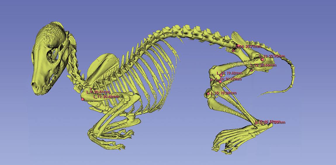
A total of four burning experiments were conducted (See Table 1). The number of experiments (n = 4) was designed to allow a preliminary assessment of reproducibility and general patterns of bone alteration, as well as to enable comparison between fleshed and defleshed/defatted bones. Prior to burning, the raccoon dog samples (1–4) were subjected to CT scanning. The CT data were analysed using the open-source software 3D Slicer to verify the post-mortem skeletal condition and to obtain baseline measurements prior to burning (See Figure 3).
The burning experiments were carried out in a medium-sized cremation furnace at an animal cremation facility (See Figure 4). The interior of the furnace consisted of a sealed combustion chamber measuring 65 × 145 cm in width and depth, and 42 cm in height. The inner walls of the chamber were lined with refractory bricks. The furnace was equipped with a kerosene-fired oil burner which automatically regulated both the air supply and fuel flow, allowing the experiments to be conducted under stable combustion conditions. The burner nozzle was located on the wall opposite the furnace entrance/exit, directing the flame into the combustion chamber. The distance between the burner nozzle and the experimental specimens was approximately 70 cm. An observation window was located on one side of the furnace, and an exhaust outlet in the ceiling. The oxidation–reduction conditions within the furnace atmosphere were not directly measured in the present study.
The furnace temperature was measured using a thermocouple installed approximately 1.3 m above the specimens, and the temperature displayed on the external panel was recorded. The recorded values represent the ambient furnace temperature and do not necessarily correspond to the effective temperature at the bone surface or within the bone. As a reference, measurements of temperature distribution in open-fire conditions indicate that, while the maximum temperature at the firebed (0 cm) reached 895°C, temperatures at the centre of the flame (15 cm above) reached 910°C, and those in the space directly above (50 cm above) reached 728°C (Japan Takibi Society, 2021). Considering these observations, the temperature at the bone surface and within the bone may have been higher than the recorded furnace temperature. Elapsed time and furnace temperature were recorded at 5–10-minute intervals. As rapid changes were observed in the early stages of burning, observations were made every five minutes, and every ten minutes in later stages, when changes became less pronounced. Changes in body position, combustion of soft tissues, bone discolouration, and fracture formation were recorded through an observation window.
After each experiment, the presence and types of discolouration, deformation and fractures were recorded. Measurable bones were subjected to osteometric analysis, and shrinkage rates were calculated by comparison with pre-burning values. Principal measurements included cranium and mandible (greatest cranial length and mandibular length) and limb bones (greatest length, mid-shaft transverse and sagittal diameters, and maximum breadth of the proximal and distal epiphyses). Measurement criteria followed established osteometric standards (von den Driesch, 1976; Murakami, et al., 1987; 1988; Nara Bunkazai Kenkyūjo Maizō Bunkazai Sentā, 2006; Gomi, 2013; 2025). In the fourth experiment, pre- and post-burning weights were recorded, and weight loss rates were calculated.
Results
Progress of Each Experiment
The progress of each experiment is shown in Appendix 1 and Table 2.
In the first experiment, a raccoon dog placed in a prone extended position (Sample 1) was burned from the caudal side. Carbonisation began in the early stage of burning, followed by the exposure of the hindlimb bones, until eventually only bones remained. In the first experiment, however, the carcass and bones were moved during burning, and therefore an accurate record of bone alterations could not be obtained. In the subsequent experiments, the specimens were not moved during burning.
In the second experiment, a raccoon dog placed in a right lateral extended position (Sample 3) was burned from the caudal side, and in the third experiment, a raccoon dog in a left lateral extended position (Sample 4) was burned from the cranial side. In both cases, carbonisation and bone exposure progressed from the side directly exposed to the flame. In the third experiment, carbonised soft tissue remained in the abdomen and hindlimbs facing away from the flame, and regional differences in bone discolouration were still evident at the end of burning.
In the fourth experiment, defleshed and defatted bones from several animal species (raccoon dog: Sample 2; pig: Samples 13 and 14; deer: Sample 15) were burned. Black discolouration first appeared in the bones closest to the flame, progressing to grey and white discolouration. Most of the bones turned white relatively early during burning. Although the temperature continued to increase after this stage, no marked change in bone colour was observed.
| Elapsed time (min after ignition) | Furnace temperature (°C) and observed changes in the carcass and bones | |||||||
| Experiment 1 | Experiment 2 | Experiment 3 | Experiment 4 | |||||
| 0 | 50 | Specimen placed in the furnace; ignition initiated. | 0 | Specimen placed in the furnace; ignition initiated. | 160 | Specimen placed in the furnace; ignition initiated. | 115 | Specimen placed in the furnace; ignition initiated. Immediately after ignition, the pig cranium and the epiphyses directly exposed to the flames blackened. |
| 5 | 556 | The skin burned and became carbonised. | 359 | The skin burned and became carbonised. Partial exposure of soft tissue observed. | 430 | The skin burned and became carbonised. Partial exposure of soft tissue observed. | 380 | Almost all bones blackened. No deformation was observed, but fractures were seen in part of the deer femoral shaft. |
| 10 | 683 | The entire body became carbonised. Skeletal elements directly exposed to the flames became visible. | 446 | The entire body became carbonised. The hindlimb bones became exposed; fractures were observed. | 498 | The entire body became carbonised. Some bones became visible. | ||
| 15 | 507 | Almost all bones turned grey to white. The deer tibial shaft fragmented. Fractures were observed in all bones. Flames erupted from the deer femur. | ||||||
| 20 | 576 | The hindlimb bones became exposed. | 509 | Exposure of the ribs and forelimb bones began. | 551 | Bones from the cranial to the forelimbs became exposed. The abdomen and hindlimbs remained carbonised. | 515 | |
| 25 | 558 | More bones became exposed. | 527 | All bones became white. | ||||
| 30 | 533 | Almost nothing but bones was left. Some carbonised soft tissue remained in places. | 538 | The fore- and hindlimb bones were almost fully exposed. The cranium and mandible exposure began. Some carbonised soft tissue remained. | 578 | Deformation and fractures were observed in the cranial and forelimb bones. The abdomen and hindlimbs remained carbonised. | 536 | |
| 35 | 487 | 545 | ||||||
| 40 | 500 | Only bones remained. | 559 | The cranium and mandible were almost fully exposed. Mainly just bones remained. | 590 | Only bones remained of the cranial and upper abdomen. The lower abdomen and hindlimbs remained carbonized. Distal hindlimb bones became exposed and showed deformation and fractures. | 553 | |
| 45 | 510 | 559 | ||||||
| 50 | 483 | Burning completed; specimen removed from the furnace. | 575 | Only bones remained. | 602 | Burning completed; specimen removed from the furnace. | 565 | |
| 55 | 572 | |||||||
| 60 | 580 | 578 | Burning completed; specimen removed from the furnace. | |||||
| 65 | ||||||||
| 70 | 586 | Burning completed; specimen removed from the furnace. | ||||||
Table 2. Experimental progress and observations. See Appendix 1 for the corresponding photographs. For each experiment (Exp. 1–4), elapsed time and furnace temperature were recorded, and changes in the carcass and bones during the burning process were observed. The experiments were conducted using a modern crematorium furnace. The temperature values, measured with a thermocouple, represent the ambient furnace temperature and not the bone surface temperature (See Figure 4). Prepared by Yasushi Furusawa.
Discolouration
As burning progressed, bone discolouration occurred stepwise.
In the first experiment, the recorded furnace temperature reached 683°C. Owing partly to movement during burning, all bones eventually turned white. By contrast, in the second and third experiments, the furnace temperature remained below 600°C, yet clear regional differences in bone colour were observed. In the second experiment, the bones on the right side directly exposed to the flame turned white, whereas grey to black discolouration persisted in the cranium, mandible and scapula on the unexposed left side (See Appendix 3: a–c). In the third experiment, the cranium, mandible and limb bones on the left side that were exposed to the flame turned white.
In contrast, bones in the abdominal and hindlimb regions where carbonised soft tissue remained showed brown to black discolouration (See Appendix 2: d). In the fourth experiment, bones turned white despite furnace temperatures remaining below 600°C (See Appendix 4).
Deformation and Fractures
The burned bones exhibited deformation, including warping, as well as several types of fractures reported in previous studies. Fractures were described following the classification proposed by Symes et al. (2015), and bending angles were roughly measured using a protractor with reference to the long axis of the bone shaft.
In the first three experiments, warping inconsistent with the original anatomical shape was repeatedly observed in the shafts of limb bones (See Appendix 2, 3). The bending angles generally ranged from approximately 20° to 40°, and the direction varied depending on bone element and side (See Appendix 2: c, f; Appendix 3: e). In the second experiment, pronounced deformation was observed in the cranium and mandible on the side exposed to the flame (See Appendix 3: a, b), while in the third experiment, marked warping occurred in the tibial shaft (See Appendix 2: h).
Fractures observed in the mandible and limb bones included longitudinal, transverse, and curved transverse fractures (See Appendix 2: a, b, enlarged view; See Appendix 3: h, enlarged view). In addition, patina fractures formed on the articular surfaces of the limb bones (See Appendix 3: h, enlarged view), while concentric ring fractures were observed on the scapula and hip bone (See Appendix 3: c, g).
The fourth experiment, meanwhile, showed different patterns. In the raccoon dog bones (Sample 2), marked warping was observed in the mandible and limb bones (See Appendix 4: a–d, f), whereas fractures were limited. In the pig bones, both deformation and fractures were minor (See Appendix 4: g–k). In the deer bones, clear warping and extensive longitudinal fractures were observed in the shafts of limb bones (See Appendix 4: l, m). Furthermore, concentric ring and curved transverse fractures formed on the articular surfaces of the tibia and calcaneus where some soft tissue remained (See Appendix 4: m, enlarged view; n).
Shrinkage
| Measurement variables and landmarks | Shrinkage rate of fleshed bones (%) | Shrinkage rate of defleshed and defatted bones (%) | ||||||||
| Raccoon dog | Raccoon dog | Pig | Deer | |||||||
| Skeletal element | Measurement variables (abbreviations) | RL | Sample 1 | Sample 3 | Sample 4 | Sample 2 | Sample 13–14 | Sample 15 | ||
| Cranium and mandible | Greatest cranial length (GL) | - | 4,7 | 9,0 | ||||||
| Mandibular length (id–C.m.) | R | 10,4 | 17,8 | |||||||
| L | 6,0 | 9,9 | 17,5 | |||||||
| Mandibular length (id–goc) | R | 9,1 | 14,4 | 6,4 | ||||||
| L | 6,5 | 8,1 | 14,8 | 9,5 | ||||||
| Humerus | Greatest length (GL) | R | 8,9 | 6,4 | 12,8 | |||||
| L | 8,3 | 6,6 | ||||||||
| Maximum breadth of the proximal epiphysis (Bp) | R | 10,6 | 8,2 | 11,3 | 21,6 | |||||
| L | 10,3 | 12,3 | 13,6 | |||||||
| Mid-shaft transverse diameter | R | 22,3 | 29,7 | 7,4 | ||||||
| L | 21,9 | 25,8 | ||||||||
| Mid-shaft Sagittal diameter | R | 20,0 | 22,1 | 9,8 | ||||||
| L | 22,2 | 22,8 | ||||||||
| Maximum breadth of the distal epiphysis (Bd) | R | 15,7 | 15,1 | 18,6 | ||||||
| L | 19,8 | 14,2 | 10,5 | |||||||
| Radius | Greatest length (GL) | R | 8,1 | 5,8 | 7,4 | 18,8 | 5,2 | |||
| L | 12,2 | 5,2 | 7,0 | |||||||
| Maximum breadth of the proximal epiphysis (Bp) | R | 15,5 | 18,5 | 18,2 | 16,8 | |||||
| L | 17,7 | 15,4 | 21,3 | |||||||
| Mid-shaft transverse diameter | R | 18,9 | 17,2 | 25,7 | 1,4 | |||||
| L | 37,3 | 15,2 | 31,3 | |||||||
| Mid-shaft Sagittal diameter | R | 16,7 | 33,9 | 38,6 | -13,4 | |||||
| L | 20,4 | 32,4 | 29,8 | |||||||
| Maximum breadth of the distal epiphysis (Bd) | R | 20,6 | ||||||||
| L | ||||||||||
| Ulna | Greatest length (GL) | R | 24,2 | 5,3 | ||||||
| L | 6,8 | |||||||||
| Mid-shaft transverse diameter | R | 0,9 | ||||||||
| Mid-shaft Sagittal diameter | R | 0,5 | ||||||||
| Distal breadth of the articular surface (DPA) | R | 26,2 | 2,3 | 16,1 | 6,8 | |||||
| L | 15,6 | 4,3 | -19,5 | |||||||
| Femur | Greatest length (GL) | R | 6,7 | 7,6 | 7,1 | |||||
| L | 11,3 | 7,8 | 7,7 | |||||||
| Maximum breadth of the proximal epiphysis (Bp) | R | 13,8 | 16,8 | 2,9 | ||||||
| L | 12,4 | 12,8 | ||||||||
| Mid-shaft transverse diameter | R | 17,0 | 16,1 | 18,4 | ||||||
| L | 14,0 | 11,4 | ||||||||
| Mid-shaft Sagittal diameter | R | 29,1 | 21,1 | 14,6 | ||||||
| L | 8,5 | 19,0 | 18,8 | |||||||
| Maximum breadth of the distal epiphysis (Bd) | R | 14,9 | 14,7 | 16,7 | 10,4 | 6,3 | ||||
| L | 14,0 | 11,3 | 15,0 | |||||||
| Tibia | Greatest length (GL) | R | 11,0 | 11,1 | 4,0 | |||||
| L | 14,5 | 10,5 | 8,9 | 6,3 | 1,8 | 7,0 | ||||
| Maximum breadth of the proximal epiphysis (Bp) | R | 19,9 | 11,7 | 12,5 | 9,3 | |||||
| L | 19,9 | 9,0 | 3,1 | 11,4 | 6,2 | 7,8 | ||||
| Mid-shaft transverse diameter | R | 24,5 | 25,6 | 6,7 | ||||||
| L | 26,8 | 24,5 | 30,9 | 4,9 | 19,7 | |||||
| Mid-shaft Sagittal diameter | R | -3,6 | 15,0 | 0,4 | ||||||
| L | 17,3 | 20,8 | -5,5 | 8,2 | 19,5 | |||||
| Maximum breadth of the distal epiphysis (Bd) | R | 4,5 | 41,0 | 19,7 | 3,9 | |||||
| L | 6,8 | 34,5 | 22,2 | 12,7 | 5,6 | 18,3 | ||||
| Fibula | Maximum breadth of the proximal epiphysis (Bp) | L | 4,4 | |||||||
| Maximum breadth of the distal epiphysis (Bd) | L | 2,5 | ||||||||
Table 3. Shrinkage rates by measurement variable. Shrinkage rates were calculated by comparing measurements obtained from 3D models prior to burning (See Figure 3) with those measured after burning. Shrinkage rates are expressed as percentages. Measurement definitions follow the osteometric methods of von den Driesch (1976), Murakami et al. (1987; 1988), Nara Bunkazai Kenkyūjo Maizō Bunkazai Sentā (2006), and Gomi (2013; 2025). Abbreviations: id = infradentale; goc = gonion caudale; C.m. = condylion mediale. Mid-shaft transverse and sagittal diameters were measured at the midpoint of the greatest length. When measurement at this point was not possible due to deformation, measurements were taken as close as possible to the midpoint. RL indicates right (R) and left (L). Prepared by Yasushi Furusawa.
Shrinkage rates for each measurement are shown in Table 3. In both fleshed and defleshed/defatted bones, shrinkage was not uniform but varied depending on anatomical region and skeletal element.
In fleshed bones, shrinkage was greater in the fore- and hindlimb bones than in the cranial bones (See Table 4, upper section). In the limb bones, shrinkage in greatest length was relatively small, whereas higher shrinkage rates were recorded for the maximum breadth of the proximal and distal epiphyses and the mid-shaft diameters, particularly in the transverse and sagittal diameters (See Table 5, upper section).
In contrast, defleshed and defatted bones showed little variation between anatomical regions (See Table 4, lower section). Shrinkage rates for each measurement item were generally uniform, and the marked regional differences observed in fleshed bones were not present (See Table 5, lower section). Interspecies differences were observed, however, in the mid-shaft transverse and sagittal diameters (See Table 3). Raccoon dog and deer showed lower values than fleshed bones, while the pig showed even lower values. Variation based on species was also observed in the greatest length shrinkage.
| Condition | Region | M | SD | n |
| Fleshed bones | Cranial bones | 7,8 | 2,0 | 7 |
| Forelimb bones | 16,3 | 10,0 | 54 | |
| Hindlimb bones | 15,4 | 9,2 | 42 | |
| Defleshed and defatted bones | Cranial bones | 12,8 | 4,1 | 7 |
| Forelimb bones | 9,8 | 9,7 | 16 | |
| Hindlimb bones | 9,9 | 5,8 | 35 |
Table 4. Comparison of shrinkage rates in cranial, forelimb, and hindlimb bones. Based on the data presented in Table 3. Upper section: fleshed bones (Sample 1, 3, and 4), lower section: defleshed and defatted bones (Sample 2, pig, and deer). Values are given as mean (M) and standard deviation (SD), and n indicates the number of measured specimens. Shrinkage rates are expressed as percentages. Cranial bones include the mandible. Prepared by Yasushi Furusawa.
| Condition | Region | Measurement variables (abbreviations) | M | SD | n |
| Fleshed bones | Limb bones | Greatest length | 8,7 | 2,4 | 19 |
| Maximum breadth of the proximal epiphysis (Bp) | 13,8 | 4,5 | 20 | ||
| Maximum breadth of the distal epiphysis (Bd) | 17,2 | 9,0 | 16 | ||
| Mid-shaft transverse diameter | 24,0 | 6,1 | 17 | ||
| Mid-shaft sagittal diameter | 20,0 | 11,2 | 18 | ||
| Defleshed and defatted bones | Limb bones | Greatest length | 9,0 | 6,2 | 12 |
| Maximum breadth of the proximal epiphysis (Bp) | 11,0 | 5,7 | 10 | ||
| Maximum breadth of the distal epiphysis (Bd) | 11,4 | 6,3 | 10 | ||
| Mid-shaft transverse diameter | 9,7 | 6,7 | 9 | ||
| Mid-shaft sagittal diameter | 8,8 | 10,7 | 9 |
Table 5. Comparison of shrinkage rates of limb bones. Based on the data presented in Table 3. Upper section: fleshed bones (Sample 1, 3, and 4), lower section: defleshed and defatted bones (Sample 2, pig, and deer). Values are given as mean (M) and standard deviation (SD), and n indicates the number of measured specimens. Shrinkage rates are expressed as percentages. Prepared by Yasushi Furusawa.
Weight Loss
In the fourth experiment, pre- and post-burning weights of defleshed and defatted bones were compared (See Table 6). Weight loss generally ranged between 40–50%, with pig bones showing the highest values, followed by raccoon dog and deer. The mean weight loss (± standard deviation) by species was 42.7 ± 5.4% in raccoon dogs, 48.7 ± 6.4% in pigs, and 40.1 ± 6.5% in deer.
By skeletal element, relatively low values were observed in the mandibles of raccoon dogs and deer, whereas higher weight loss was recorded in the scapula of raccoon dogs and in the ulna, radius, and tibia of pigs; however, the number of specimens for pigs and deer was limited.
| Element | RL | Weight loss (%) | ||
| Raccoon dog (Sample 2) | Pig (Sample 13–14) | Deer (Sample 15) | ||
| Cranium | - | 38,4 | ||
| Mandible | - | 30,8 | 31,9 | |
| Humerus | R | 39,6 | ||
| L | ||||
| Radius | R | 43,5 | 56,9 | |
| L | ||||
| Ulna | R | 42,8 | 52,5 | |
| L | ||||
| Scapula | R | 52,2 | ||
| Sacrum | - | 45,0 | ||
| Hip bone | R | 47,4 | ||
| L | 34,1 | |||
| Femur | R | 43,4 | 40,6 | |
| L | 44,6 | |||
| Tibia | R | 43,9 | ||
| L | 45,2 | 50,8 | 47,7 | |
| Fibula | R | |||
| L | 45,1 | |||
| M | 42,7 | 48,7 | 40,1 | |
| SD | 5,4 | 6,4 | 6,5 | |
| n | 12 | 5 | 3 | |
Table 6. Weight loss rates of defleshed and defatted bones. Weight loss was calculated as the percentage reduction from pre-burning to post-burning weight. Weight loss rates are expressed as percentages. Values are given as mean (M) and standard deviation (SD); n indicates the number of measured specimens. RL indicates right (R) and left (L). Prepared by Yasushi Furusawa.
Discussion
Overview of the Experimental Results
This study examined how differences in burning conditions influence bone discolouration, deformation and fracture formation, shrinkage, and weight loss through burning experiments using a modern cremation furnace. The results confirmed that the presence or absence of soft tissue, the body position and the flame exposure direction affect bone alteration. They additionally suggested that burning temperature and duration have a certain influence on bone discolouration and shrinkage. These findings provide basic data for assessing burning conditions based on the observation of burned bones. The results are discussed in detail below.
Presence or Absence of Soft Tissue and Bone Alteration
Effects on Discolouration
Bone discolouration during burning has traditionally been understood mainly in relation to burning temperature (Shipman, et al., 1984). Recent studies, however, have indicated that the relationship between temperature and discolouration is not necessarily straightforward (Ellingham, et al., 2015; Rosa, et al., 2023; Galloway, et al., 2024). Furthermore, it has been reported that prolonged burning at relatively low temperatures may produce discolouration and chemical alterations similar to those observed at higher temperatures (Gallo, et al., 2023).
In the second and third experiments of the present study, in which the recorded furnace temperature remained below 600°C, only the areas directly exposed to the flame showed whitening, whereas grey, brown and black colouration persisted in non-exposed areas. By contrast, in the fourth experiment, where defleshed and defatted bones were burned, almost all bones turned white despite furnace temperatures also remaining below 600°C. These results indicate that discolouration is determined not only by temperature and burning duration but also by multiple additional factors, including flame exposure conditions and the presence or absence of soft tissue. In particular, soft tissue has been suggested to delay whitening by partially shielding bone surfaces from heat (Symes, et al., 2015; Gonçalves, 2025), and the results of the present study are consistent with this interpretation.
Effects on Deformation and Fracture Formation
In the experimental burning of carcasses, both deformation and fractures were pronounced. Differences in body position and flame exposure direction produced regional and lateral differences in the degree and direction of bending, suggesting that heat distribution contributes to the formation of bending. The observed fractures were mainly longitudinal and transverse fractures, although curved transverse fractures were found around muscle attachment areas of the limb bones, as well as the scapula and hip bone. In burned human bodies, heat-induced muscle contraction is known to cause flexion of the limbs (the so-called pugilistic posture) and the formation of curved transverse fractures (Symes, et al., 2015, pp. 34–36). Similar phenomena have also been reported in animals (Wohlsein, et al., 2016, pp.1009–1010; Carroll and Smith, 2018). The present results also support the possibility that thermal muscle contraction contributes to the formation of curved transverse fractures. On the other hand, damage to joint regions, which are generally considered vulnerable to thermal destruction, was not frequently observed in the present experiments.
In contrast, defleshed and defatted bones showed relatively limited fracture formation but exhibited pronounced deformation and extensive longitudinal fractures. This pattern does not necessarily correspond with previous studies, which suggest that deformation rarely occurs in dry bones (Whyte, 2001). Yet, the experimental conditions of the present study resulted in the observation of a different tendency, possibly influenced by the degree of defleshing and defatting, bone tissue structure and heating conditions.
Effects on Shrinkage
Previous studies have reported that shrinkage tends to be greater in cross-sectional diameters and epiphyseal breadths than in bone length (Hummel and Schutkowski, 1986; Thompson, 2005). In the present experiments involving fleshed bones, shrinkage was similarly greater in the mid-shaft transverse and sagittal diameters, and in the maximum breadth of the proximal and distal epiphyses than in the greatest length. This result is consistent with the authors’ hypothesis regarding the radius from Pit C316 at the Dobashi site, interpreted to be the result of cremation, in which shrinkage in the mid-shaft diameters was greater than in the greatest length (Furusawa and Nara, 2025).
In comparison, no marked differences were observed between the shrinkage rates of greatest length, mid-shaft diameters and epiphyseal breadths in defleshed and defatted bones. This may be related to the prior loss of organic components, although the result does not directly correspond to the weight-loss results discussed below. Furthermore, the pig samples included limb bones that may derive from a juvenile individual, as indicated by unfused epiphyses. It is known that limb bones of juvenile individuals exhibit bone structures different from those of adults (König and Liebich, 2016, pp.36–44), and the observed differences in shrinkage rates may reflect the developmental stage. The defleshed and defatted bones used in this study, moreover, may differ in properties from archaeological dry bones that have undergone burial, and this factor should be taken into account when interpreting the results.
These results indicate that the presence or absence of soft tissue has a significant influence on bone alteration, and that it can be assessed to a certain extent through macroscopic observation. At the same time, the experiments also suggest that these effects vary depending on combinations with other factors, including burning temperature and duration, body condition, anatomical region, bone tissue structure, animal species and development stage. Further investigation is therefore required to clarify these interrelations.
Effects of Body Position and Flame Exposure Direction
In the second and third experiments, the combination of body position and flame exposure direction produced clear regional and lateral differences in both discolouration and shrinkage.
In the second experiment, in which the body was placed in a right lateral position and exposed to flame from the caudal side, whitening occurred on the right side, while black colouration persisted on the left side. Conversely, when, in the third experiment, the body was placed in a left lateral position and exposed to flame from the cranial side, the flame-exposed left side whitened earlier. In contrast, the thick, soft tissues of the abdomen acted as a thermal shield, delaying whitening in the abdominal and hindlimb regions. A similar trend was observed in shrinkage rates, with higher shrinkage recorded on the side directly exposed to the flame (See Table 7). Additionally, in the measurements of fore- and hindlimb bones (See Table 3), many variables showed higher shrinkage values on the right side in the second experiment and on the left side in the third experiment, confirming lateral differences corresponding to body position.
| Cranial bones | Forelimb bones | Hindlimb bones | |
| Sample 1 | - | 17,6 | 14,2 |
| Sample 3 | 7,3 | 15,2 | 16,7 |
| Sample 4 | 9,0 | 16,5 | 14,6 |
Table 7. Differences in shrinkage rates depending on the flame exposure direction. Based on the data presented in Table 3. Cranial bones include the mandible. The number of measurements used for the calculations was n = 7 for cranial bones, 54 for forelimb bones, and 42 for hindlimb bones. Shrinkage rates are expressed as percentages. The flame exposure direction varied between specimens (caudal-side exposure in Sample 3 and cranial-side exposure in Sample 4). Sample 1 is included for reference. Prepared by Yasushi Furusawa.
Chatzikonstantinou (2022) has reported that direct flame exposure enhances bone fragmentation, while Garriga (2023) has noted that skeletal elements located closer to the fire source tend to show lower survival rates. These studies indicate that the spatial relationship between bones and fire source influences heat distribution. The results of the present experiments further suggest that the combination of body position and flame exposure direction also plays a major role in determining heat distribution.
Burning Temperature, Burning Duration, and Bone Alteration
Although the present experiments could not establish a clear quantitative relationship between effective bone surface temperature and burning duration, they did confirm the influence of relative thermal load, whereby bones directly exposed to higher-temperature flames showed greater shrinkage. It should be noted, however, that the temperatures recorded in this study represent the ambient temperature inside the furnace and may not necessarily correspond to the effective temperature at the bone surface or within the bone.
The rate of weight loss in the defleshed and defatted bones was higher than that reported in previous studies (Trotter and Peterson, 1962). Because flames were observed erupting from within the bones during burning, it is possible that a certain amount of organic components, particularly moisture and lipids, remained within the bones. The degree of defatting and air-drying treatment may therefore have influenced the results. These results are generally consistent with existing models in which the weight of burned bones decreases through stages of moisture loss and loss of organic components (Imaizumi, et al., 2014; van Hoesel, et al., 2019), although the relationship between weight loss and shrinkage still requires further investigation.
Implications for Archaeological Burned Bones
The results of this study provide important insights for interpreting the burning process of the human remains from Pit C316 at the Dobashi site and Pit SK439 at the Kamino site.
The shrinkage, deformation, and characteristic fracture patterns observed in the burned human bones from Pit C316 at the Dobashi site are most consistent with Hypothesis 1, suggesting that they were cremated at a high temperature within a relatively short period after death. In addition, the lateral and regional differences observed in discolouration and deformation may reflect variations in thermal load that different parts of the skeleton were exposed to and may be explained by variations in body position and flame exposure direction as proposed in Hypothesis 2.
By contrast, the burned human bones from the Kamino site’s Pit SK439 show mixed patterns of discolouration, deformation and fractures, and differences in the presence or absence of bioerosion have also been observed. These characteristics may be explained by a combination of Hypotheses 1–3. In other words, the evidence suggests that at least two different burning processes may have been practiced: cremation carried out shortly after death, and secondary burning of bones following primary burial.
The results of this study provide a new perspective for understanding the diversity of burning practices in Jōmon funerary systems, and present experimental baseline data for estimating burning conditions based on combinations of bone discolouration, deformation, fractures and shrinkage.
Limitations of the Experiment and Future Research
This study is a preliminary experiment conducted using a modern crematorium furnace, and the furnace structure as well as the burning conditions do not necessarily correspond to those of prehistoric open-air burning. In addition, there were certain limitations regarding the condition of the specimens, temperature measurement, and sample size.
In future research, it will be necessary to verify the present results in open and heterogeneous thermal environments, such as through open-air burning experiments, to directly measure the temperature distribution within and around the bones, incorporate a wider variety of skeletal materials, and integrate these approaches with histological analyses, in order to test and supplement the findings of this study and improve the accuracy of reconstructing burning processes.
Conclusion
This study experimentally examined how burning conditions - including the presence or absence of soft tissue, body position, flame exposure direction, burning temperature, and burning duration - affect bone alteration. Although the experimental conditions do not account for all possible variables, the results nevertheless demonstrate that these factors influence bone alteration and help to clarify the relationships between burning conditions and patterns of discolouration, deformation, fractures, shrinkage, and weight loss. These patterns provide useful observational criteria and analytical indicators for investigating burning processes.
The findings of this study provide baseline data for designing future comparative burning experiments and refining interpretative models of archaeological burned human remains. In particular, they offer a useful reference framework for examining differences between burned human remains from the Dobashi and Kamino sites.
Acknowledgements
The authors would like to express their sincere gratitude to all those who provided valuable guidance and support throughout this study and the preparation of this manuscript.
We are particularly grateful to Annika Wagner, Hiroki Tsuruta, Satoko Kumaki, Mara Schmidt-Falbe, Motoyasu Kato, Junmei Sawada, Yasuo Hagihara, Aiko Saso, Fumiko Saeki, and Koji Tatsumi for their assistance and support.
We also thank the staff of the Pet Funeral Service Centre, Focal Co., Ltd., the Niigata Prefecture Archeological Research Corporation, and Mitsuke City Office.
Conflict of interest
The authors declare no conflicts of interest.
Country
- Japan
Bibliography
Abe, Y., 2009. Jōmon jidai iseki ni okeru shōkotsu seisei no jikken kōkogakuteki kenkyū [Experimental archaeological study of burned bone formation at Jōmon period sites]. Sundai Shigaku, 136, pp. 31–55
Carroll, E.L. and Smith, M., 2018. Burning questions: investigations using field experimentation of different patterns of change to bone in accidental vs deliberate burning scenarios. Journal of Archaeological Science: Reports, 20, pp. 952–963. https://doi.org/10.1016/j.jasrep.2018.02.001
Chatzikonstantinou, Y., 2022. The use of fire in the manipulation of the deceased through experimental approaches. In: M. Mărgărit and A. Boroneanț, eds. Recreating artefacts and ancient skills: from experiment to interpretation. Cetatea de Scaun, pp.307–324
Chatzikonstantinou, I., Fülöp, K., Chronaki, S., Snoeck, C., Kiriatzi, E. and Triantaphyllou, S., 2025. Experimental insights into different funerary burning conditions on Crete Island, Greece. Journal of Archaeological Science: Reports, 62, pp.105067. https://doi.org/10.1016/j.jasrep.2025.105067
Correia, P.M., 1997. Fire modification of bone: a review of the literature. In: W.D. Haglund and M.H. Sorg, eds. Forensic taphonomy: the post-mortem fate of human remains. CRC Press, pp.257–293
Ellingham, S.T.D., Thompson, T.J.U., Islam, M. and Taylor, G., 2015. Estimating temperature exposure of burnt bone—A methodological review. Science and Justice, 55, pp.181–188. https://doi.org/10.1016/j.scijus.2014.12.002
Furusawa, Y. and Nara, T., 2025. Fundamental research of burned human bones: a case analysis of the C316 burned human bones from the Dobashi site, Niigata Prefecture, Japan. Anthropological Science (Japanese Series), 133(1), pp.1–13. https://doi.org/10.1537/asj.250503
Gallo, G., Ushakov, S.V., Navrotsky, A. and Stahlschmidt, M.C., 2023. Impact of prolonged heating on the color and crystallinity of bone. Archaeological and Anthropological Sciences, 15, pp.143–167. https://doi.org/10.1007/s12520-023-01842-0
Galloway, A., Pope, E. and Juarez, C., 2024. Bone color changes and interpretation of the temperature/duration of fire exposure to human remains in the forensic context. WIREs Forensic Science, 6(4), e1517. https://doi.org/10.1002/wfs2.1517
Garriga, J.A., 2023. Skeletal alteration of burnt remains through fire exposure. In: S. Ellingham, J. Adserias-Garriga, S.C. Zapico and D.H. Ubelaker, eds. Burnt human remains: recovery, analysis, and interpretation. Hoboken, NJ: Wiley, pp.113–132. https://doi.org/10.1002/9781119682691.ch7
Gomi, Y., 2013. Inu no tōgai shishikotsu keisoku ni tsuite [On the measurement of canine cranial and limb bones]. Dōbutsu Kōkogaku, 30, pp.197–220
Gomi, Y., 2025. Hosoku inu no tōgai shishikotsu keisoku ni tsuite [Supplement: on the measurement of canine cranial and limb bones]. Dōbutsu Kōkogaku, 42, pp.63–64
Gonçalves, D., 2025. The effect of soft tissues in burnt human skeletal remains. International Journal of Osteoarchaeology, 35, pp.383–391. https://doi.org/10.1002/oa.70024
Hummel, S. and Schutkowski, H., 1986. Das Verhalten von Knochengewebe unter dem Einfluss höherer Temperaturen: Bedeutungen für die Leichenbranddiagnose [The behaviour of bone tissue under the influence of high temperatures: implications for the diagnosis of cremated remains]. Zeitschrift für Morphologische Anthropologie, 77, pp.1–9
Imaizumi, K., Taniguchi, K. and Ogawa, Y., 2014. DNA survival and physical and histological properties of heat-induced alterations in burnt bones. International Journal of Legal Medicine, 128(3), pp.439–446. https://doi.org/10.1007/s00414-014-0988-y
Japan Takibi Society, 2021. Temperature of bonfires. (research report). Available at: < https://takibisociety.web.fc2.com > [Accessed: 1 April 2026]
Kato, M. and Kato, M.Y., 2025. Niigata-ken Murakami-shi Kamino iseki no chōsa kenkyū hōkoku (1) [Report on the archaeological investigation of the Kamino site, Murakami City, Niigata Prefecture (1)]. Kenkyū Kiyō, 15, pp.9–33
Kato, M.Y., 2025. Jōmon jidai shōjinkotsu ichiranhyō [Catalogue of burned human remains from the Jōmon period]. In: Nihon Kōkogaku Kyōkai 2025-nendo Niigata Taikai Jikkō Iinkai, ed. Essa kara mita rettō no kōryū to chiiki shakai, pp.207–218
König, H.E. and Liebich, H.-G., 2016. Karā atorasu jūi kaibōgaku, jōkan (zōho kaitei dai 2-han) [Color atlas of veterinary anatomy, volume 1 (revised and expanded 2nd edition)]. Tōkyō: Midori Shobō
Murakami, Y., Hirata, H. and Okuzaki, M., 1987. Tanuki, Nyctereutes procyonoides viverrinus kokkaku no gensunzu to sokuteichi I. Tōgaikotsu [Life-size skeletal drawings and measurements of the raccoon dog (Nyctereutes procyonoides viverrinus), Part I: skull]. Joshi Eiyō Daigaku Kiyō, 18, pp.149–162
Murakami, Y., Hirata, H. and Okuzaki, M., 1988. Tanuki, Nyctereutes procyonoides viverrinus kokkaku no gensunzu to sokuteichi II. Tsuikotsu, rokkotsu, zen-shikotsu, kankotsu, kōshikotsu [Life-size skeletal drawings and measurements of the raccoon dog (Nyctereutes procyonoides viverrinus), Part II: vertebrae, ribs, forelimb bones, hip bone and hindlimb bones]. Joshi Eiyō Daigaku Kiyō, 19, pp.145–159
Nara Bunkazai Kenkyūjo Maizō Bunkazai Sentā, 2006. Dōbutsu kōkogaku no tebiki. Nara: Nara Bunkazai Kenkyūjo Maizō Bunkazai Sentā
Nara, T., Saeki, F. and Furusawa, Y., 2023. Fuhen: shōjinkotsu ni tsuite [Appendix: on burned human remains]. In: Agano-shi Kyōiku Iinkai, ed. Dobashi iseki: Agano-shi maizō bunkazai hakkutsu chōsa hōkokusho, 19, pp.1–36
Nara, T., 2026. Jinruigaku ni miru shōjinkotsu [Burned human remains from an anthropological perspective]. In: Niigata-ken Maizō Bunkazai Sentā, ed. Hakkutsu! Niigata no iseki 2025. Niigata: Niigata-ken Maizō Bunkazai Sentā, pp.16–17
Osaka, D. and Yoneda, M., 2025. Jōmonjin wa itsu yakareta no ka? [When were Jōmon people burned?]. In: Nihon Kōkogaku Kyōkai 2025-nendo Niigata Taikai Jikkō Iinkai, ed. Essa kara mita rettō no kōryū to chiiki shakai, pp.192–195
Rosa, J., Marques, M.P.M., Gonçalves, D. and Ferreira, M.T., 2023. Half a century of systematic research on heat-induced colour changes in bone – a review. Science and Justice, 63(5), pp.573–580. https://doi.org/10.1016/j.scijus.2023.07.002
Shipman, P., Foster, G. and Schoeninger, M., 1984. Burnt bones and teeth: an experimental study of color, morphology, crystal structure and shrinkage. Journal of Archaeological Science, 11(4), pp.307–325. https://doi.org/10.1016/0305-4403(84)90013-X
Snoeck, C. and Schulting, R.J., 2013. Fire and bone: an experimental study of cremation. EXARC Journal, 2013(2). Available at: https://exarc.net/ark:/88735/10111.
Symes, S.A., Christopher, W.R., Chapman, E.N., Gipson, D.R. and Piper, A.L., 2015. Patterned thermal destruction in a forensic setting. In: C.W. Schmidt and S.A. Symes, eds. The analysis of burned human remains, 2nd ed. Academic Press, pp.17–59. https://doi.org/10.1016/B978-0-12-800451-7.00002-4
Tomioka, N., Katagiri, A. and Adachi, N., 2014. Miyagi-ken Sendai-shi Dōnokuchi iseki shutsudo no chūsei kasōkotsu no kenkyū [Study of medieval cremated human remains excavated from the Dōnokuchi site, Sendai City, Miyagi Prefecture]. Handayama Chiri Kōko, 2, pp.79–90
Thompson, T.J.U., 2004. Recent advances in the study of burned bone and their implications for forensic anthropology. Forensic Science International, 146S, pp.S203–S205. https://doi.org/10.1016/j.forsciint.2004.09.063
Thompson, T.J.U., 2005. Heat-induced dimensional changes in bone and their consequences for forensic anthropology. Journal of Forensic Sciences, 50(5), pp.1008–1015. https://doi.org/10.1520/JFS2004297
Trotter, M. and Peterson, B.R., 1962. The relationship of ash weight and organic weight of human skeletons. Journal of Bone and Joint Surgery, 44-A(4), pp.669–681
van Hoesel, A., Reidsma, F.H., van Os, B.J.H., Megens, L. and Braadbaart, F., 2019. Combusted bone: physical and chemical changes of bone during laboratory simulated heating under oxidising conditions and their relevance for the study of ancient fire use. Journal of Archaeological Science : Reports, 28, pp.102033. https://doi.org/10.1016/j.jasrep.2019.102033
von den Driesch, A., 1976. A guide to the measurement of animal bones from archaeological sites. Peabody Museum Bulletin 1. Harvard University
Whyte, T.R., 2001. Distinguishing remains of human cremations from burned animal bones. Journal of Field Archaeology, 28(3–4), pp.437–448. Available at: https://www.jstor.org/stable/3181445
Wohlsein, P., Peters, M., Schulze, C. and Baumgärtner, M., 2016. Thermal injuries in veterinary forensic pathology. Veterinary Pathology, 53(5), pp.1001–1017. https://doi.org/10.1177/0300985816643368